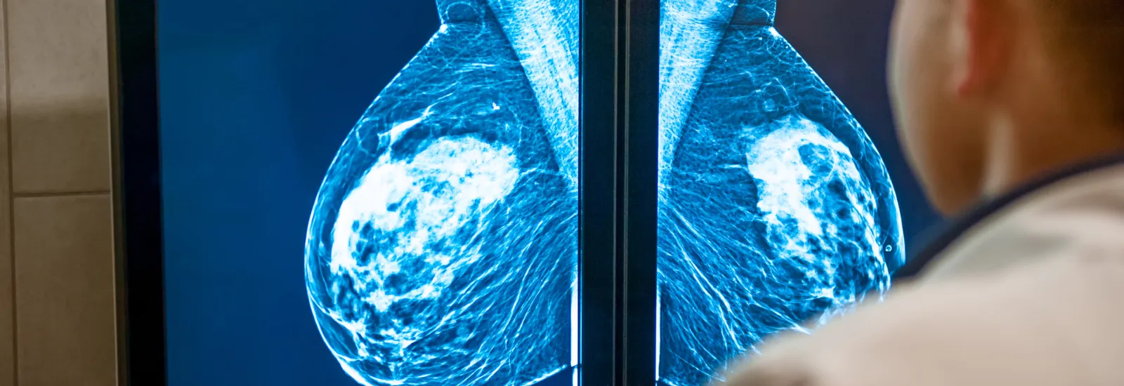
Mammograms: When you get called back
Maybe you've heard this over and
over again: Early detection saves lives. So you follow your provider's advice
and schedule your annual mammograms. Only this time, you're called in for
follow-up tests. Should you fear the worst? Definitely NOT. Among women
who get called back for additional breast testing, the American Cancer Society says fewer than 10% receive a
cancer diagnosis.
All call backs are not created equal
There are several reasons you could
get called in for a follow-up mammogram, including:
- The images of your breasts may show an area the radiologist would like to see more clearly or in a different projection.
- An area in the breast looks different from the previous year there are many reasons why one part of the breast may appear different. Follow-up images can uncover the cause.
- A mass or other suspicious area is present in one or both breasts. Before moving to more invasive diagnostic techniques, follow-up imaging can help determine whether anything additional needs to be done.
If you haven't had one already, your
provider may recommend a 3D mammogram that gives a more in-depth view of your breasts.
Risk
factors for repeat mammograms
Although anyone can get called back
for repeat mammograms, some people are at higher risk of receiving an abnormal
result.
Those risk factors include:
- Having your first mammogram. Your provider has nothing to compare the results of your first mammogram to.
- You haven't reached menopause. Callback mammograms are more common among younger women.
- Your breasts are dense. Dense breasts are more common if you're on hormone replacement therapy and are not overweight. Younger women are more likely to have dense breasts, but women of any age can have them too.
Diagnostic tests after the initial mammogram
If you're called back after a
screening mammogram, additional imaging is often the first step forward. These
are diagnostic mammograms, but you will go through the same process as your
annual test. In most cases, a diagnostic mammogram is all you need to
determine all is well.
In other cases, your provider may
want you to have additional testing, such as:
Breast
ultrasound. A
breast ultrasound is the same technology used to visualize babies in pregnant
women. For this noninvasive test, the sonographer spreads gel on the skin and
uses a wand to take images of your breast. As it travels, the wand produces
sound waves, which create a real-time view of the breast's interior. These
images help determine whether the area is suspicious and needs further testing,
such as a biopsy.
Breast
biopsy. If an abnormality
is found on the mammogram, a Breast Center nurse will refer you to a general
surgeon to determine the best approach needed for your situation. During a
breast biopsy, a small tissue sample is removed for examination. This tissue is
often taken with a thin needle, guided by ultrasound or another imaging
technique. Once your surgeon has the sample, it is sent to the laboratory for
testing. This will lead to a definitive diagnosis.
After your mammogram
Many follow-up breast imaging and
biopsy results show no cancer. If cancer is present, your provider will explain
the stage and grade of your cancer and discuss the best treatment options for
you.
Remember, early
detection means early treatment and a greater chance of survival. If cancer is
detected in the breast area only and hasn't spread, the five-year survival rate is 99%. Routine screening is so important — and it all starts with a high-quality
mammogram.
Looking for accurate, convenient
mammography services? Schedule your advanced 3D mammogram today at Reid Health Breast Center.

